- Mallomonas
- a - b
- Mallomonas acaroides
- Mallomonas acaroides var. inermis
- Mallomonas actinoloma
- Mallomonas actinoloma var. maramuresensis
- Mallomonas actinoloma var. nadiensis
- Mallomonas adamas
- Mallomonas akrokomos
- Mallomonas alata
- Mallomonas alata f. hualvensis
- Mallomonas allorgei
- Mallomonas alphaphora
- Mallomonas alpina
- Mallomonas alveolata
- Mallomonas annulata
- Mallomonas areolata
- Mallomonas asmundiae
- Mallomonas bangladeshica
- c - d
- Mallomonas calceolus
- Mallomonas canina
- Mallomonas caudata
- Mallomonas clavus
- Mallomonas conspersa
- Mallomonas corcontica
- Mallomonas corymbosa
- Mallomonas costata
- Mallomonas crassisquama
- Mallomonas crassisquama var. papillosa
- Mallomonas cratis
- Mallomonas cristata
- Mallomonas cucullata
- Mallomonas cyathellata
- Mallomonas doignonii
- e - h
- i - l
- m - o
- pa - pi
- Mallomonas palaestrica
- Mallomonas paludosa
- Mallomonas papillosa
- Mallomonas papillosa var. monilifera
- Mallomonas parvula
- Mallomonas paxillata
- Mallomonas peronoides
- Mallomonas phasma
- Mallomonas pillula f. exannulata
- Mallomonas pillula f. latimarginalis
- Mallomonas pillula f. pillula
- Mallomonas pillula f. valdiviana
- po - pu
- r - s
- t - z
- a - b
- Synura
- Paraphysomonas
- Spiniferomonas
- Chrysosphaerella
- Chrysodidymus
- Polylepidomonas
Paraphysomonas
The genus Paraphysomonas comprises solitary, colourless flagellates, spherical to slightly oval cells, covered with siliceous spine scales and/or basket or plate scales. The cells bear two flagella of unequal length. They are usually found as freely swimming cells, but they also occur attached to bacterial mats or other substrates via a stalk at the posterior end of the cell.
Paraphysomonas species feed on various different organisms, such as bacteria, diatoms or small chlorococcalean algae. The prey is ingested at the base of the flagella after being pulled in via the motion of the long flagellum, the food is incorporated into vacuoles within a few seconds. Undigested particles are extruded around the posterior end of the cell.
Identification of Paraphysomonas species is based on the ultrastructure of the silica scales. The scale structures are various; open mesh structures, basket scales, perforated plate and basket scales as well as the common plain spine with an oval to round base. Of the ~56 species of Paraphysomonas not all species have just one scale type, many have two forms of scales per cell, and in varying numbers. Scales are developed within vesicles that are intimately associated with cisternae of rough endoplasmic reticulum in the peripheral cytoplasm.
![]() |
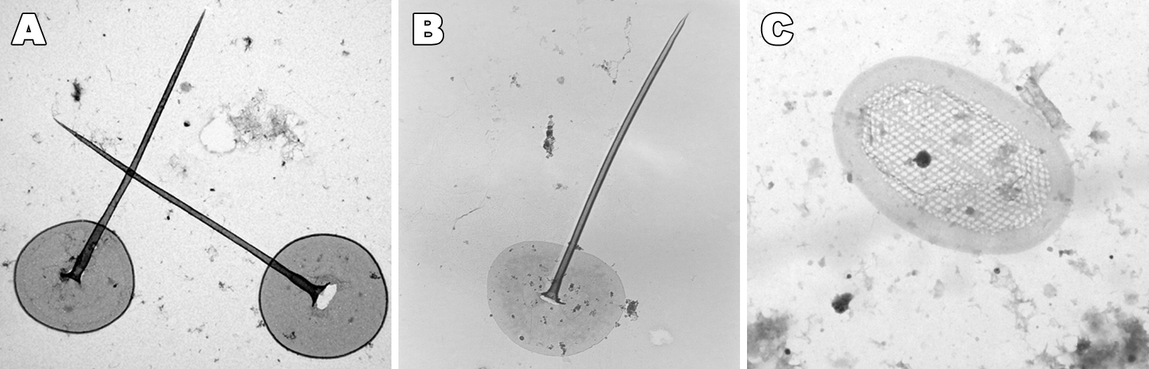
| Paraphysomonas siliceous structures. A. Circular flat disc scale with a centrally protruding tapering spine and thickened rim (P. vestita morphotype) B. Circular flat disc scale with a central spine (P. imperforata). C. Eliptical, perforated plate scale with a central spine one of the forms is has (P. takahashii). D. Bowl-shaped, elliptical scale with a wide rim (P. punctata). E. Complex, perforated, three-dimensional crown scales (P. cf. butcheri). F. Formation of two, morphologically different scales in one cell (Paraphysomonas sp.) |

